Harvard Medical School - Department of Neurobiology - Corey Lab
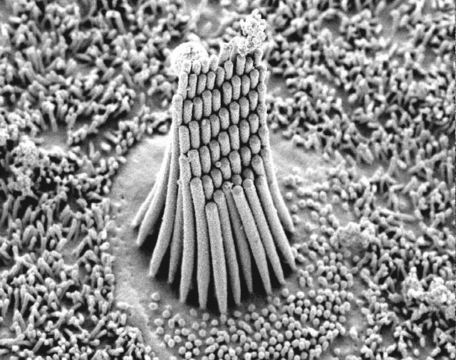
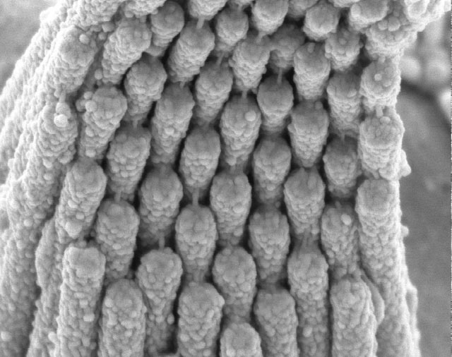
While we marvel at discoveries in technology and materials, SEM images like these of the inner ear remind us how living organisms have used the trial-and-error engineering of evolution to develop amazing structures. The SEM images show the hair-like projections, known as stereocilia, that extend from the top of each of the thousands of receptor cells in the inner ear. They are connected by "tip links," fine filaments that are tensioned by sound and that pull open ion channels. Such channels are the gates for electric current; in the ear their opening converts sound into neural signals. Studying the inner ear's receptor cells is the passion and life's work of Dr. David P. Corey of the
Howard Hughes Medical Institute and Department of Neurobiology, Harvard Medical School, Boston.
 Harvard Medical School - Department of Neurobiology - Corey LabCategory
1990s image of the inner ear at 12,000X
 Harvard Medical School - Department of Neurobiology - Corey LabCategory
1990s image of the inner ear at 45,000X reveals tip
 Harvard Medical School - Department of Neurobiology - Corey LabCategory
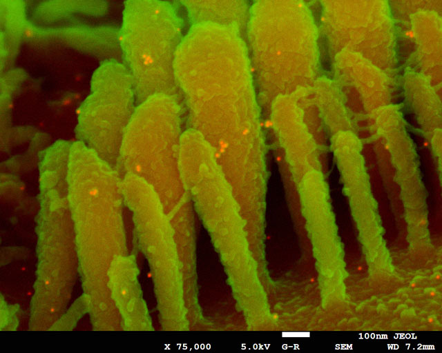
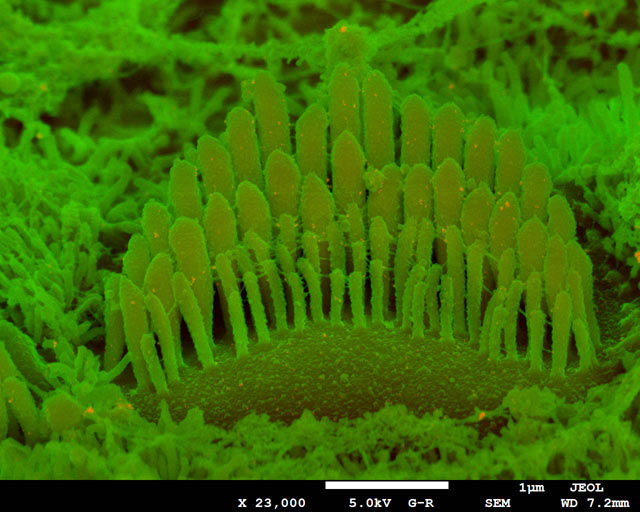
2012 FE SEM images (false colored) of the hair cell of the inner ear
 TitleCategory
2012 FE SEM images (false colored) of the hair cell of the inner ear
"We study these channels in hair cells -- the receptor cells of the inner ear, which are sensitive to sounds or accelerations," he explains. Hair cells are a third the diameter of a human hair and the stereocilia are ~300 nm wide, less than the wavelength of light. The tip links, at 10 nm diameter, can barely be seen in SEM.
Corey collaborated with JEOL in 1991 to observe tip links with the then-new field emission SEM, showing for the first time that experimental manipulations that broke tip links abolished sensitivity to sound, and thus demonstrating their critical role in hearing. "We used FESEM because the tip links are so small that 30 kV electrons go right through them. With field emission we could get good images down around 5 kV."
Back then, he says, "We knew about the biophysics and structure but had no idea of the molecular components. Now that protein candidates for the transduction apparatus are being identified, it is critical to localize them to within a few nanometers." Corey and his colleagues are now back at JEOL, using backscatter imaging for immuno-gold labels. Scientists at JEOL are helping them to devise the best procedures for sample preparation for optimum imaging.
Ultimately the research helps to understand hearing loss, especially inherited disorders that affect proteins of the transduction complex. One, for instance, is Usher Syndrome. "It's a nasty disease" Corey says, "where a child is born completely deaf and then as a teen loses vision as well. It's pretty devastating." Studying the tip link structure reveals something about how to address this genetic defect, but also helps scientists understand how noise trauma can cause hearing loss in normal individuals. Loud noise, such as extremely loud music or an explosive blast, can break these tip links. "Without tip links the cells simply don't work. Luckily the tip links can be repaired within 5-10 hours, but if the exposure is continued the hair cell eventually dies. It's an incredibly fragile structure."