Howard Hughes Medical Institute – Electron Microscopist Lita Duraine
Electron Microscopist Lita Duraine seated at the JEOL JEM-1400Plus TEM in the Bellen Lab.
As a certified Electron Microscopy Specialist at Howard Hughes Medical Institute (HHMI), Lita Duraine enjoys a unique perspective by helping investigators in the field of medical and biological discoveries. Scientists, graduate students, post-docs, and collaborators from outside her lab rely on her expertise to produce perfectly detailed images that allow them to better understand muscular and neurological systems. She has primarily used the Transmission Electron Microscope (TEM) during her time at HHMI and also at previous organizations to explore the intricate details of a fruit fly’s (Drosophila melanogaster) physiology and the inner ear of a toad fish, all to help advance human medical research in such areas as ALS, Parkinson’s, anti-gravitational studies, and even the use of stents.
Lita has been working at HHMI for nearly seven years now, preparing samples and taking pristine images for her clients, including 25 students during a semester, in the Bellen Lab, under the direction of
Dr. Hugo Bellen.
“I help everyone with their samples - from dissection, tissue processing and embedding, sectioning and TEM examination. I have had the opportunity to work on a wide diversity of specimens in my career from plants, mammals, fish, reptiles, and now insects,” she says.
“Manipulating Drosophila melanogaster is a bit challenging from an electron microscopy point of view, but is so indispensible for genomic research in Amyotrophic lateral sclerosis (ALS), Alzheimer’s, and other debilitating diseases.”
She marvels at how the common fruit fly, so different from humans, actually shares many things in common and serves as a model organism for studying genetics and other fields of research. “It’s amazing how in nature everything interacts and touches each other. We can look at the muscular system of drosophila as larvae to study ALS in humans. We can also look at the drosophila retina system in the brain for neurological studies in humans. The homology is similar.”
 Howard Hughes Medical InstituteCategory
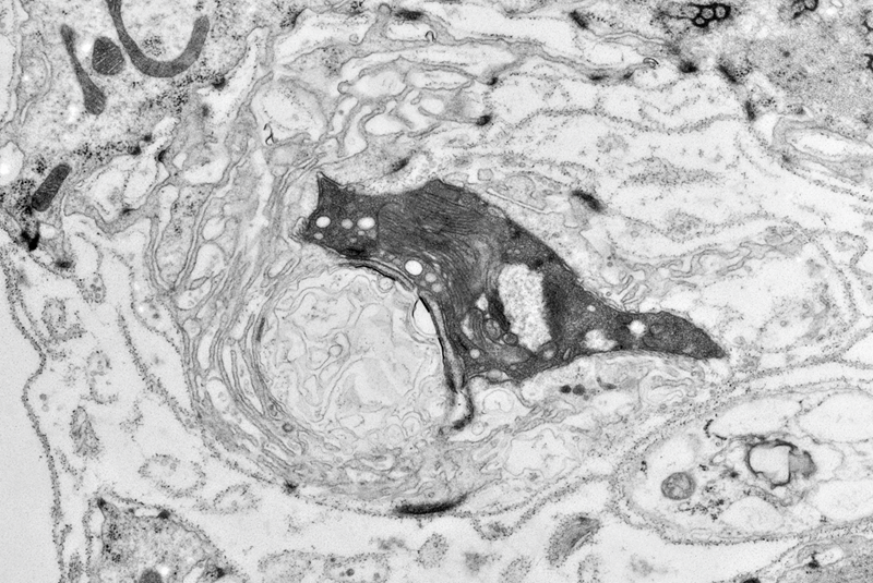
2017 Image Contest Winner “Cat and Ball” Drosophila melanogaster neuromuscular junction with bouton.
 Howard Hughes Medical InstituteCategory
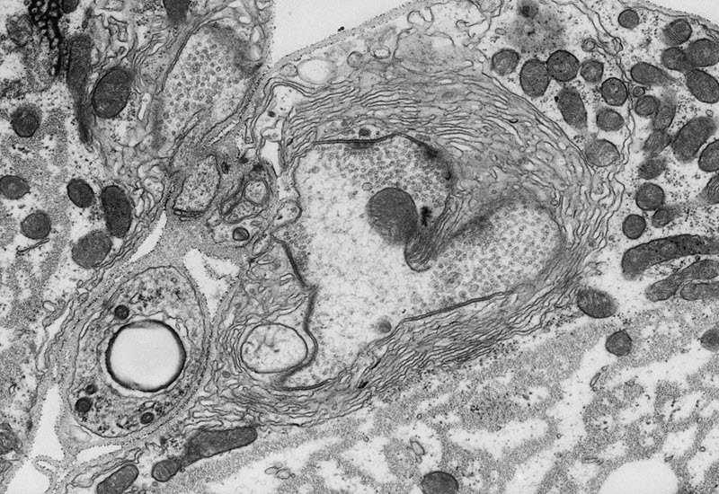
2018 Image Contest Winner “Love Me, Love My Bouton” - Drosophila larvae were fixed in Modified Karnovski’s fix, osmicated, dehydrated in graded ethanols, and embedded in Embed 812. Tissue was cut on a Leica EM UC7 Ultramicrotome.
Microscopy is a satisfying career that began later in life for Lita. She graduated from San Joaquin Delta College in California in 2001. The field was not one she had known much about until some life changes made her reconsider her path towards a pharmacology degree.
“It wasn’t like I always knew I wanted to be an electron microscopist,” she states. Earlier interests included exploring the chemistry of making natural cosmetic products and the intricacy of making jewelry. “I knew I loved science and gravitated towards that by studying pharmacy.” Part way through her degree, she learned about the electron microscopy program at San Joaquin Delta College. It sparked a new passion that became, well, very focused, so she enrolled and never looked back.
She studied both biology and crystalline materials microscopy at the college when it was just building the microscopy center. She was taught by Dr. Judy Murphy in the earlier days of the program. It was an invigorating time for her, and opened many doors.
She started her new career and was soon setting up SEM and TEM labs at NASA Ames Research Center and Boston Scientific, as well as teaching others to operate the suite of instruments. She also consulted in the set up and teaching at other labs. In 2007 she moved from California to Texas, when she joined the Howard Hughes Medical Institute.
HHMI in Houston is connected with Baylor College of Medicine, but is essentially a separate entity with 350 Principal Investigators throughout the world.
Huda Y. Zoghbi, MD, is the director of the Jan and Dan Duncan Neurological Research Institute at Texas Children’s Hospital, of which the Bellen Lab is a part.
The variety of studies has captured her imagination. “That’s the fun part, you learn through a lot of different projects.” One day she may be looking at retina samples and the next day brain, heart, or muscle samples.
 Howard Hughes Medical InstituteCategory
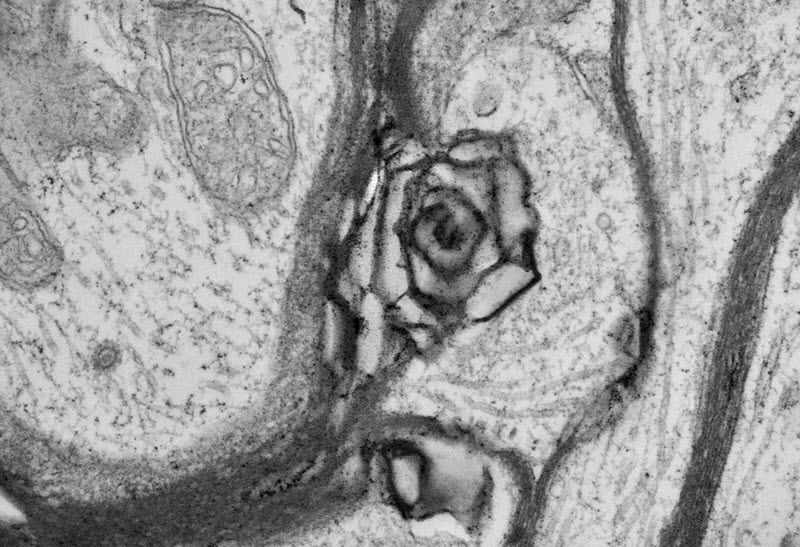
“Axon Rose” The image is a representation from genotype VPS11 from drosophila brain tissue. VPS11 is one of many Vacuolar Protein Sorting genes that we studied in our lab. The image shows a mutant axon gone awry amongst other brain cells.
 Howard Hughes Medical InstituteCategory
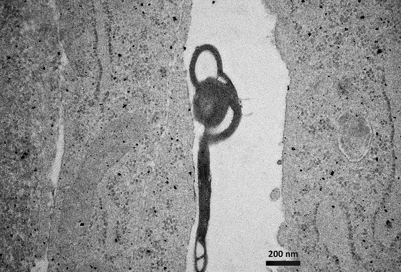
“Hallowed Vacuole” - Image of mutant human fibroblast cells with a large vacuole. Found this guy hanging around putting a halo on his head. Micron bar represents 200 nm, total magnification is 20,000X. Cells were plated, scraped and processed with Modified Karnovski’s fix and Osmium, dehydrated with graded ethanols, and embedded in Embed 812 resin.dehydrated in graded ethanols, and embedded in Embed 812. Tissue was cut on a Leica EM UC7 Ultramicrotome.
In addition to actually taking the images, sample preparation is something she excels at. It’s a process that is carefully done, from using uranyl acetate and lead citrate stain, a two-part process for conductivity, usually at room temperature. “In my experience it’s better, because what happens when you put cold water on tissue? It kind of shrivels up. I’m old-fashioned, I don’t cut and stain on the same day and I don’t stain and view in the TEM on the same day. Each step is done on a different day – there’s not as much outgassing of heavy salts in the TEM that will build up.”
She keeps a calendar and careful records as she works on several clients’ projects simultaneously. Once samples are loaded into the sample holder in a grid, with as many as 10 sections on a slot grid, imaging goes quickly. “Just bring up the TEM and condition the grid, focus the image and get it in alignment, you can pretty much roll through and take 10-20 images – I average a micrograph every minute or less with the JEOL 1400+ TEM.”
Some of her pictures have been chosen as winners of the JEOL image contest. Getting beautiful images after just a few seconds is what keeps Lita so happy with the
JEM-1400 TEM, not to mention the service contract that keeps things running smoothly.
Her techniques have allowed her to easily retrieve precise information on each image and section, and she can even go back and look at grids that are as much as five years old. “I keep everything! It’s a Lita rule, don’t throw anything away.”
While she runs the TEM so effortlessly, she is also as knowledgeable of the Scanning Electron Microscope thanks to her training with Delta College. “I’ve met many who do microscopy who didn’t have that schooling and are very good at what they do, but schooling allowed me to learn more techniques and details than I otherwise would have. People I knew in school had come from other scientific backgrounds and then made a choice to go this way, or others just wanted a career in microscopy.” Being a certified electron microscopist is a specialized field but it allows one to be an integral part of, and a valuable contributor to, research and discovery.
“If you love detail, if you love intricacy and like working on tiny things, it’s very rewarding to get to see a side of nature and creation that you do not get to see anywhere else. It leaves you with some questions answered and sometimes you’ll have even more. It’s a rewarding field and I’m glad I was led to it,” says Lita.