Snowflakes, Superglue, and SEM
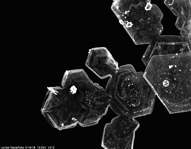
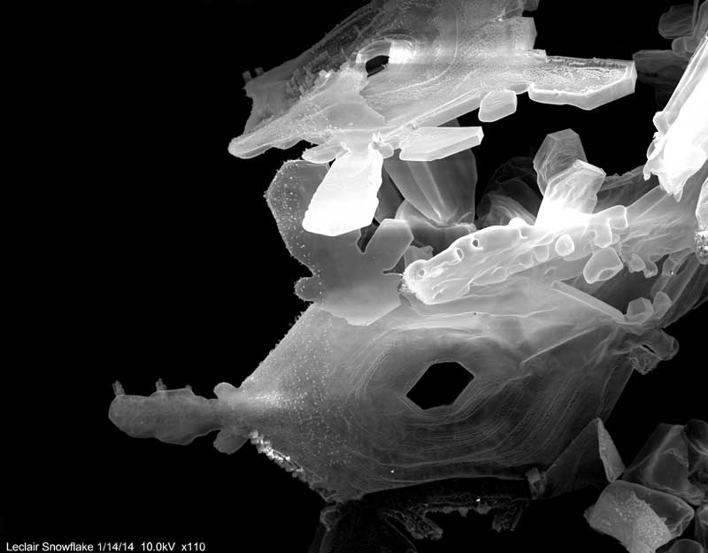
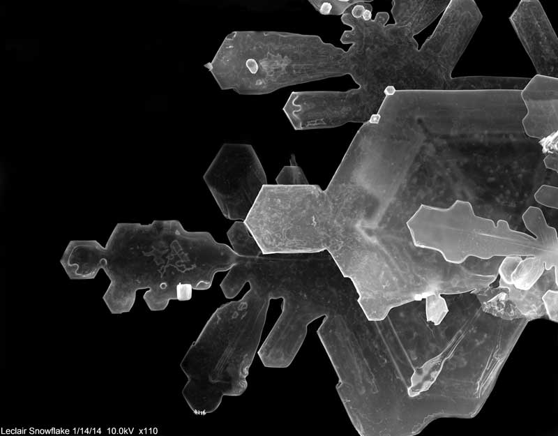
Intrigued by cryo microscopy, and even further inspired when his professor mentioned that one could take SEM images of ice crystals in snow, an undergraduate biotechnology student was determined to capture his own images of snowflakes. He had two problems, however. There is no cryoSEM in the lab, and the freeze fracture system is in need of repair.
"I decided if it could be done on a cryo, there had to be a way to do it on an older microscope," says Mike LeClair, who is a senior at
SUNY College of Environmental Science and Forestry (SUNY-ESF) in Syracuse, NY. He wanted to add snowflake images to the portfolio he is making this year while using the school's 18-year-old JEOL JSM-5800LV. His professor, Robert Smith, suggested he look for older papers on the topic, which turned out to be hard to find.
As the snow began to fall on the campus, Mike decided to contact JEOL directly. He reached Mark Melvin, Sales Engineering, who asked Asst. Director and applications specialist Donna Guarrera for advice. She referred him to a webpage -
snowcrystals.com - for a detailed technique that uses superglue to create a "fossil" or replica of the snow crystal. He followed the procedure, but was pretty sure the effort had failed. "The stubs that I put the glue on were a mess. They dried into big clumps on top and no individual flakes were preserved." Three weeks later, however, he took another look at his samples and was surprised to find "perfect little preserved snowflakes that still glittered like snow would in the sun." He found that the excess glue had pooled around the base of the stubs while it dried. The snow that had accumulated there, instead of on the tops of the stubs, turned out to be the perfect samples. And then, his first images were so exciting he quickly let us know the experiment had worked.
 SUNY College of Environmental Science and Forestry (SUNY-ESF)Category
Snowflakes, Superglue, and SEM
 SUNY College of Environmental Science and Forestry (SUNY-ESF)Category
Snowflakes, Superglue, and SEM
 SUNY College of Environmental Science and Forestry (SUNY-ESF)Category
Snowflakes, Superglue, and SEM
"I am definitely trying it again; I actually have a sample case sitting outside in the snowstorm right now," he reported in January. "This time I left the stubs out of the equation and just put glue directly in the bottom of the sample holder." He's hoping to get intact flakes this time, without damage from wind or jostling the sample case in his pocket. After he waits for the glue to dry.
Mike will complete his SEM course and then start his TEM course this spring as he completes his biotechnology degree with a minor in microscopy. His goal is to work in a molecular biology lab and it would be even better if that position required use of the EM.
"Microscopy is something that, until this fall, I had never considered as a career, but the possibility is definitely there." His portfolio to date includes his best results in imaging a multitude of samples, but his success with snowflakes has him thoroughly enjoying the process of solving the mystery of how to image something with a few extra challenges to consider.
Dr. Susan Anagnost, Director of the Center, and Professor Robert Smith, describe the microscopy program: "Because we have a very diverse curriculum here at ESF and we often partner with Syracuse University and Upstate Medical University, our samples could range from bacteria to wood to insects to graphene nanoparticles. Sometimes, the project director would like images from light, scanning and transmission on the same subject. Taxonomic studies on insects for instance are easier to accomplish on the SEM than the light microscope! One of the most challenging studies was a project involving thin sectioning TEM of rubber tire components. A very recent and exciting study is currently being conducted to trace certain proteins involved in stem cell communication with neighboring cells by immune-electron microscopy and TEM.
At our NC Brown Center for Ultrastructure Studies, each machine is used to teach undergraduate and graduate students the techniques necessary to obtain Journal quality images. They are critical instructional components for our Microscopy Minor, taught in the SCME Department at ESF. Those courses are: SEM and TEM (2 to 5 credit hours), Fundamentals of Microscopy (3 credits), TEM use for nanoparticle studies (2 credits) and Medical and Industrial Applications of Electron Microscopy (3 credits). Last year, we had over 70 students enrolled in these courses. For many graduate students, expertise in electron microscopy is fundamental to their research projects. Our outside users normally ask us to investigate a proprietary company project, involving SEM or TEM, for which we charge a fee. Over the years, our center has investigated scores of outside projects for local and national companies. Our capabilities are listed on our
web site.
Our students are from SUNY-ESF and Syracuse University and come from a variety of departments: Physics, Engineering, biomaterials science, Environmental Science, Biology, Biotechnology, Environmental Engineering, Polymer chemistry and Paper Science and others."
Working with this older equipment -- the SEM was purchased 15 years ago and the TEM nearly 30 years ago -- poses no issues in terms of user friendliness and the laboratory experience. Students follow exercises that build on one another, "so that by the end of the semester, the student is able to prepare and image a sample of their choosing and incorporate it into a picture portfolio for class evaluation."
Aganost and Smith look forward to obtaining new TEM capabilities in the near future that would allow them to "delve deeper into nanoparticle research, identify and map atoms involved in structural events, view frozen images of cellulose and the molecular motor that winds it into place. With STEM the would be able to observe delicate samples that we feel could be valuable assets in drug delivery. Disease vectoring insects could be studied to determine if metal-bearing proteases are involved in transport of disease to salivary glands for re-infection."