Universidad de la República - Uruguay
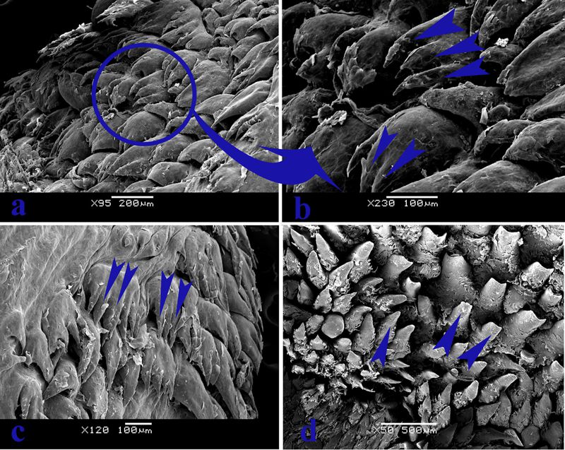
CW from left: SEM image of equine gutteral pouch; Prof. William Perez; Denton Vacuum drying apparatus; JEOL JSM-5900LV Scanning Electron Microscope, Science Faculty building
“Anatomy is to physiology as geography is to history; it describes the theatre of events.”
― Jean Fernel French physician who introduced the term "physiology" to describe the study of the body's function.
The study of anatomy is the foundation for understanding the form and structure of tissues, muscles, and bones in the body, and how those structures adapt to function. Scientists have studied anatomy for centuries, but in the last 50 years high resolution, high magnification electron microscopy and advanced imaging techniques have allowed them to come to a deeper understanding.
Since 2000, scientists from the University of the Republic (UdelaR) in Uruguay have been revealing remarkable details about anatomy that has had an impact on research and veterinary medicine worldwide, with numerous publications to their credit.
Currently, Dr. Silvia Villar Arias (Faculty of Science, UdelaR) and Dr. William Perez (Professor of Anatomy at the Veterinary Faculty) are developing comparative anatomy studies, focusing on the native fauna of Uruguay. According to Dr. Villar, "Anatomy is an exciting discipline at the confluence of evolutionary convergence and genetic adaptation how it expresses the development of pathological processes due to wildlife exposure to pollutants from agro-industrial origin. That has led us to obtain promising results, several of which have been published or are in press. We are now developing projects spanning diverse disciplines such as evolution, anatomy, histology and genetic toxicology."
This work is very exciting for Dr. Perez, who says, "The anatomical knowledge of many animal species is very scarce. The generation of such information is vital for later studies of other disciplines related to the conservation of species or their exploitation. Most of our work has been centered in the digestive, reproductive, cardiovascular and musculoskeletal systems. The idea is to conduct future research on the nervous system at the microscopic and ultrastructural level. The anatomical studies are performed by simple dissection or using stereoscopic microscope, photography, basic measurements, histology and various types of microscopy (confocal, fluorescence, SEM and TEM)."
Dr. Villar adds, "The biological sciences within microscopy are, in my view, the most exciting because the development of protocols and treatments for more reliable, artifact-free results are very challenging. Together with other researchers, I have worked on projects related to new treatments for celiac disease, the study of certain types of sclerosis and its relation to the reorganization of the extracellular matrix of the myelin sheaths, the study of host-bacteria interaction in mammals and plants, as well as cellular and genetic impact analysis of the interaction of pathogens with urinary tract cells. I have also had the opportunity to interact with scientists whose research is related to the area of materials, working in the analysis of ash for end deposition, corrosion processes of industrial piping, studies of geological samples, particles, material and nanostructures related to graphene for the development of supercapacitors.
 Universidad de la República - UruguayCategory
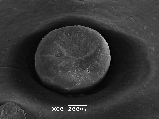
Gustative fungiform papillae of fallow deer (Dama dama) tongue.
 Universidad de la República - UruguayCategory
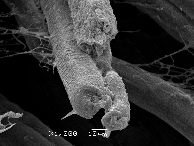
Fibers of retractor bulbi muscle of the eye of southern Minke whale (Balaenoptera bonaerensis).
 Universidad de la República - UruguayCategory
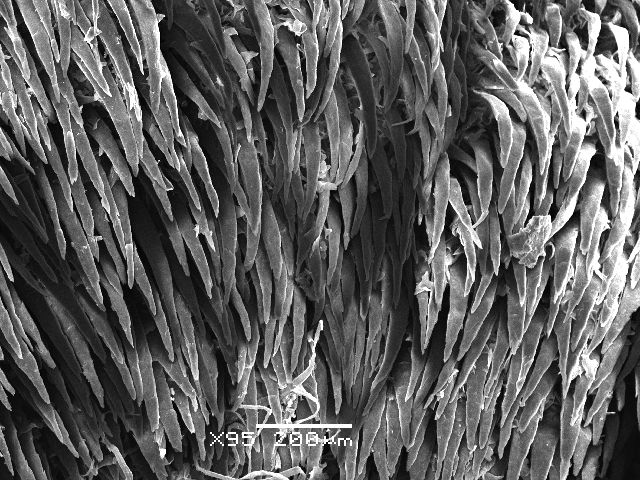
Ctenomys pearsoni tongue.
 Universidad de la República - UruguayCategory
"Training students in the last five years has been unprecedented in this laboratory, both in the area of environmental toxicology and epifluorescence as well as in scanning electron microscopy," Dr. Villar adds.
Dr. Perez has coauthored papers with fellow researchers across the globe, including Serkan Erdogan, Department of Anatomy, Faculty of Veterinary Medicine, Namık Kemal University, Tekirdağ, Turkey who has worked on anatomical and SEM studies of such topics as the tongue and lingual papillae of the meerkat, comparing the different information on the morphology of the other carnivore species, and also the tongue of the pampas deer. Recently, with Horst König University of Veterinary Medicine in Vienna, author of several texts on anatomy of domestic animals, Perez has studied the gutteral pouch of the horse.The Uruguay group have some interesting insights.In the equids (horse family) and some species such as the desert hyrax. The guttural pouch is one of a pair of chambers in the neck just behind the skull and below the ears. These pouches are on both sides of the head and are air-filled out-pouchings or evaginations to the Eustachian tube, leading into the nasopharynx. The aim of this work for morphological characterization of these complex structures is important because they exhibit of pathologies, like mycosis, empyema, and infections. In the grooves you can find bacteria, fungi and so on,it is just a refuge for microorganisms. There are very few studies of this type of organ done with the SEM and they are very excited about these findings. Equine research will continue to be a topic as because there are many strange cases with neurological implications and paralysis to be investigated.
Dr. Silvia Villar Arias and Dr. William Perez use the JEOL JSM-5900LV Scanning Electron Microscope which was installed in 2000. Dr. Arias learned SEM as an undergraduate student, analyzing evolutionary processes linked to reorganization of genetic material. For a while her work on environmental pollution and its impact on cell activity and genetic material steered her towards fluorescence microscopy. Since 2008 she has integrated the SEM in her work. Dr. Perez began using SEM in 2013.
The work is also exciting for the country of Uruguay, which was colonized by the europeans and gained independence in the early 1800s. In 2013 it was named "Country of the Year" by The Economist. The University of the Republic, founded in 1843, is the country's most important, oldest, and largest university.
For Further Information
Bibliography