August 31, 2010 (Peabody, Mass.) -- JEOL USA, a leading supplier of high resolution Transmission Electron Microscopes (TEMs) for biological and materials research, announces that Emory University in Atlanta, Georgia, has selected two JEOL TEMs for the Robert P. Apkarian Integrated Electron Microscopy Core (RPAIEMC).
The two TEMs, one operating at 120kV and the other at 200kV, will be used in Life and Soft Materials Sciences research. As such, the instruments are equipped for imaging of conventionally obtained stained samples or sections as well as frozen hydrated specimens. State-of-the-art software will allow for high throughput using techniques ranging from tomography to single particle imaging. RPAIEMC staff, members of the Wright lab, and users from the region will image and study a broad range of biological and soft materials samples such as: infectious viruses; pathogenic and nonpathogenic bacteria; mammalian tissues; self-assembled peptide matrices; and nanoprobes.
Use of Phase Plate Technology Increases Imaging Contrast for Biological Structures
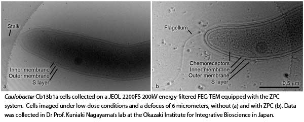
The JEOL model JEM-2200FS TEM with its in-column energy filter and thin film/electro-static phase plate technology will be the showpiece of the expanded EM laboratory. “Phase plate technology, exclusively available commercially for TEMs through JEOL, increases biological specimen imaging contrast by a factor of 3-5, which is going to be of tremendous benefit for imaging a range of cryo-specimens from single particles all the way to sectioned materials,” said Dr. Jaap Brink, JEOL’s TEM Product Manager
Emory to Serve as Unique Center for Biological Imaging
“This will be the first biological Field Emission TEM in the state of Georgia and will establish Emory University as a unique center for biological imaging. The 2200FS will be dedicated to cryo-imaging of biological and soft materials specimens,” said Dr. Elizabeth R. Wright, Director of the RPAIEMC and Assistant Professor in the Department of Pediatrics, Emory University School of Medicine, and Georgia Research Alliance Distinguished Investigator. Dr. Wright is the Principal Investigator for the major research instrumentation grant awarded by the National Science Foundation (NSF: 0923395), which supported the acquisition of the 200 kV FEG-TEM. As part of the evaluation process, Dr. Wright collected data on bacterial samples using phase plates specifically designed for the JEOL TEM by Prof. Kuniaki Nagayama’s lab at the Okazaki Institute for Integrative Bioscience in Japan, the lab that developed the technology. She also collected data in the lab of Dr. Wah Chiu at the National Center for Macromolecular Imaging at Baylor College of Medicine in Texas, a center for excellence in cryo-electron microscopy.
Practical Use of Phase Plate Technology for Higher Contrast TEM Imaging
The practical use of phase plate technology provides a marked increase in specimen contrast not typically seen in biological samples imaged under cryo-conditions. “My lab specializes in cryo-EM and cryo-electron tomography (cryo-ET) of bacteria and viruses. We do not alter the structure of the sample through the addition of contrast enhancing stains. Therefore, the primary way we obtain greater contrast is to apply a defocus to the image. However, this is at the expense of resolution. The use of a phase plate, both the Zernike type and the electro-static type, will allow us to maximize the contrast of unstained samples without the use of a defocus and thus retain the resolution. It is an amazing benefit to have this technology to apply to structural studies of pathogens,” Wright said. Dr. Wright studies the basic structure of several pathogenic viruses and bacteria in order to develop novel vaccines and therapeutics. Her research involves examining viruses, such as HIV-1, measles, and respiratory syncytial virus (RSV), which are generally100 to 300 nm in size. Specific targets include the examination of viral assembly and maturation, as well as the viral glycoproteins that attach to and fuse viruses to the target cell.

Collaborative Partnership to Advance Phase Plate Technology
A collaborative partnership between Emory University and JEOL USA, aimed at maximizing the efficacy of the phase plate technology, will enhance the visibility of the RPAIEMC throughout the EM community and will enable JEOL to favorably present the latest and most sophisticated cryo-TEM in a state-of-the-art facility.
Dedicated TEM for Conventional Samples
A JEOL model JEM-1400 was purchased simultaneously with support from a shared instrumentation grant from the National Institutes of Health (NIH: 1S10RR025679-01), which was awarded to Prof. Paul Spearman, Nahmias-Schinazi Research Professor and Vice Chair for Research Department of Pediatrics, Emory University and Chief Research Officer, Children’s Healthcare of Atlanta. The RPAIEMC at Emory provides regional investigators with instrumentation, training, and expertise in all areas of biological and soft materials electron microscopy. The new 120 keV TEM will be used for imaging thousands of research samples and Wright says it “will be dedicated primarily to conventional EM of sectioned and negatively stained materials. It will also allow users to perform basic tomography on many sample types.”